Original Article
Nerve root sparing in posterior approach corpectomy and thoracolumbar spine reconstruction using an expandable cage: A novel technique
1 Department of Orthopedic Surgery, Soroka University Medical Centre and Faculty of Health Sciences, Ben Gurion University of the Negev, Beer-Sheva, Israel
2 Independent Physician, Institute of Pathology, Soroka University Medical Centre and Faculty of Health Sciences, Ben Gurion University of the Negev, Beer-Sheva, Israel
Address correspondence to:
Nissim Ohana
Institute of Pathology, Soroka University Medical Centre 1, Rager Boulevard, P.O. Box 151 ,
Pin- 84101, Israel
Message to Corresponding Author
Article ID: 100002VNP05NO2017
doi: 10.5348/VNP05-2017-2-OA-1
Access full text article on other devices

Access PDF of article on other devices

How to cite this article
Ohana N, Benharroch D, Sheinis D. Nerve root sparing in posterior approach corpectomy and thoracolumbar spine reconstruction using an expandable cage: A novel technique. Video J Orthop Surg 2017;2:1–5.Abstract
Aims: Thoracolumbar fractures, especially when disintegration of the bony fragments occur, require surgery. However, no specific operative approach has so far been given precedence. We herewith propose a variant surgical procedure which may be indicated for the above purpose.
Methods: The suggested technique should be carried out in a single stage, by posterior approach only. Following the generation of an appropriate access and evacuation of the bone debris, a titanium mesh cage should be inserted, using an original manipulation, shown in the enclosed video and which will cause minimal or no damage to the neural elements.
Results: Fourteen patients were submitted to our variant operation, seven with Type A (according to the Aospine classification) fracture (three with two simultaneous fractures). Three suffered from C type fractures; Most patients presented with a preserved neurological status; Three were paraplegic on admission (Frankel grade A). The most marking benefits of this technique included optimal height restoration, immediate stability, and direct canal decompression. All patients except one improved in the postoperative period, by at least one grade on Frankel scale.
Conclusion: The proposed procedure variant fulfilled most of our expectations, to the benefit of the patients. In addition, the duration of the surgery was shorter than that of the A-P procedure. Our technique could be further extended to fractures above and below the T11-L2 segment, and also include patients with Frankel grade A lesions.
Keywords: Burst fracture, Mesh cage insertion, Nerve roots, Surgery, Thoracolumbar spine
Introduction
Injuries to the spinal column are not rare, a proportion being related to a blunt trauma. In the US, about 160,000 individuals suffer from such injuries yearly. Of these, about 20% are limited to the thoracolumbar (TL) region (T11-L2) of the spine, the most vulnerable area being localized to the L1 vertebra [1].
While considering the treatment options, most of the orthopedic surgeons favor surgery, performed for neurological decompression, or in order to restore the alignment, but also to preserve a sagittal balance [2].
The available surgical techniques for unstable burst fractures (classes A3 or A4 according to the AOSpine classification), include either a posterior access, especially for indirect canal decompression and fusion. This is performed by fusion of a long segment (three vertebrae above the lesion and two below it), to prevent kyphosis, or by fusion of a shorter segment, if the vertebral body is only mildly collapsed. An alternative technique is utilized for anterior conus compression and in cases of severe vertebral body comminution. This option is also used for a canal compression of 70% or more, or for a degree of kyphosis superior to 30o [3],[4].
Finally, a combined anterior and posterior access is also employed for an unstable burst fracture. This provides for immediate stability, complete kyphosis correction, together with its maintenance, as well as for complete spinal canal decompression [5],[6].
As discussed the presently available treatment options, two questions are put forward regarding fractures of the TL vertebrae. First, what is the most adequate classification system for surgical or non-surgical decision making? Moreover, for a TL burst fracture with an incomplete neurological deficit, what are the optimal surgical approach and stabilization techniques? [7],[8],[9].
To try and answer the above questions it has been stated that there is no specific surgical approach in the case of a TL burst fracture with incomplete neurologic damage that has any advantage, so far as neurologic recovery is concerned [10],[11].
We therefore propose a variant surgical procedure - partial vertebral body replacement for complex spinal fractures encompassing: a posterior approach; direct canal decompression with vertebral body replacement; anterior column reconstruction and posterior instrumented arthrodesis.
Materials and Methods
Using a posterior access method, the affected spinal segment is exposed. As a preparatory step for fixation, pedicle screws are introduced into the vertebrae above and below the fracture.
At first, laminectomy of the involved vertebra is performed. Excision of the facet joint, together with the adjacent pedicle, is carried out. As a rule, this stage can be achieved only on one side (the more compressed or more comminuted) of the involved vertebra. Once access to the vertebral body is obtained, a direct, unilateral decompression of the spinal canal is engaged until a partial corpectomy is completed. The tip introduced herein is first to abrade and scrub the caudal and rostral endplates from all soft tissue and cartilage present. At this point, the titanium mesh cage should be inserted into the vertebral body cavity. As a rule, this step entails the sacrifice of one or two pertinent nerve roots. However, this is not tolerable at the level of the lumbar spine, since the nerve roots function at this level is vital. A means of preserving the nerve roots at any level along the T-L spine is presented: a mesh cage of appropriate size is chosen, and is maintained at a collapsed state. To prevent injuries to the nerve roots during its insertion, the mesh cage is initially placed onto the corpectomy site, perpendicularly to the long spinal axis, and then introduced into the bone cavity, using a 90° rotary movement (Video 1). This maneuver, performed with a 90? long clamp, uses the cavity at the apex to stir the cage. Once the collapsed cage rests within the corpectomy site, the elongation device is carefully attached to it and allows the cage expansion into the vertebral body bone cavity.
Results
Ours was a retrospective four-year study at the Rabin Medical Center, Petah-Tikva, Israel, on complex TL fractures with variable neurological status. We review 14 patients, eight males and six females, mean age 30 (range 16-48). Seven patients had a type A fracture (according to the AOSpine classification) (1–A2.2; 2–A3; 4–A4), of which 3 patients had 2 simultaneous fractures (1–A4+C1; 2–A4+A2).
The time elapsed between injury and surgery varied from 3–20 hours and was mostly within 7 hours.
As mentioned in the methods, 14 patients were operated on, using our variant procedure, although they did not conform strictly with the original criteria of inclusion: they suffered from: 1–B2.1; 1–C1.3; 1–C3.1 and 1–C3.2 fractures, including dislocation of the spine.
The neurological status did not comply for all patients with the original definition, either, as three patients presented with Frankel grade A on admission. However, two of these improved to grade D and one to grade E, within 30 days of surgery. Most of the remainder improved by one grade, within this time lapse. Only one patient regressed by one grade on Frankel scale, from D to C, but we have not been able to determine the cause of this mild deterioration in neurological status.
The sagittal profile, as defined by mean preoperative lordosis was 18o. Operative improvement will result in a mean lordosis of 32o.
The most marking benefits from our recommended surgical technique included immediate stability, optimal height restoration and direct canal decompression. All patients were allowed full mobilization once their medical status permitted.
One patient developed a perioperative infection that was treated with irrigation and intravenous antibiotics. There was no need for instrumentation removal. We have mentioned above the patient with a limited neurological deterioration at follow-up. No mortality was observed.
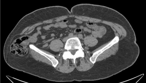
A 16-year-old female was admitted to our Institute following a suicide attempt by jumping from the third floor. On admission she was alert and hemodynamically stable. She could not feel or move both her lower extremities and both her feet were deformed. On examination she was ASIA A at the level of D12. No bowel or bladder function was evident. A significant deformation of both feet was later diagnosed as comminuted fractures of ankles and feet bilaterally. Imaging studies (Figure 1) showed severe burst fractures at two levels: at L1 and L4. Three hours after admission, the patient was operated on and had partial vertebral bodies resection through posterior approach of L1 and L4, using our root sparing technique. The corpectomies were followed by posterior spinal fusion from T11 to S1 (Figure 2). After completion of her spine surgery, the patient was submitted to external fixation of both her ankles and feet. Following recovery in the intensive care unit, and short delay in the orthopedic department, the patient was transferred to the rehabilitation department.
During the recovery period, the patient regained partially but not effective movements of her lower extremities. Before transfer to rehabilitation, she was assessed as ASIA C. Later, the patient slowly regained her ability to stand and walk with crutches. One year following surgery she was able to stand and walk with one cane and was able to control her bowel and bladder functions. Her feet progressed to partial function only. Serial radiographs showed at that stage solid and stable arthrodesis of the lumbosacral spine. A computed tomography scan demonstrated optimal decompression of the spinal canal.
The clinical data of the remaining patients in our study were summarized graphically. Figure 3 depicts the distribution of the fractures along the spine. The majority of the fractures was found between D12 and L3.
The neurological status, at presentation and during progression, using the Frankel score, is depicted in Figure 4. Only 11 patients are summarized in this figure, and all, except one show progression towards Frankel grade E.


Discussion
Spinal fractures are not rare. The main location is in the TL region (T11-L2) in about one-fifth of the cases. When considering burst fractures of the TL area only, several intervention techniques are available, but none is consistently considered the favorite. The injury classification and damage severity score, previously proposed, have not been proven conclusive [12],[13]. They might not, therefore, represent the basis of a consensual treatment modality. We have suggested a surgical procedure based on a one stage posterior approach with no need for nerve root sacrifice [14]. This method includes posterior access, which nevertheless allows for complete evacuation of the bony fragments from the vertebral body as well as from the spinal canal, when necessary.
A special feature is documented in the video sequence: this is the modus operandi through which the titanium mesh cage is introduced into the vertebral body bone cavity, in a way that will not damage the nerve roots [15],[16] (Video 1).
The advantages of the procedure encompass direct canal decompression, optimal height restoration and immediate stability acquisition. Additional benefits include a shorter surgical procedure, as compared with a stage AP technique. Moreover, except for one patient whose neurologic status regressed, from an unknown reason, most patients improved in the perioperative period at least by one grade on Frankel scale. Three patients progressed from grade A to grade D (two patients) and to grade E (one patient). Suzuki et al. [17], employed a comparable posterior surgical technique for burst fractures of the lumbar spine for his patients, except for the following: they have shortened the lumbar spine and used simple, not expandable cage. This led to subsidence to a lower level and to kyphosis of the affected segment. Nevertheless, Suzuki et al. have suggested what we urge to emphasize herein: this approach is feasible and can even be improved on if our technique is adopted.
It is of note, that although originally meant to treat patients with a burst fracture of the T-L (D11-L2) spine and with incomplete neurological deficit, we have expanded the spectrum of fractures that we operated on, using the proposed method of surgery, to include 14 patients. Among these, we also report on patients with Frankel grade A; with fractures above D11 and below L2; as well as a few with C types fractures with dislocation of the spine. All obtained a clear advantage from surgery, using our proposed variant procedure.
References
1.
Katsuura Y, Osborn JM, Cason GW. The epidemiology of thoracolumbar trauma: A meta-analysis. J Orthop 2016 Jul 21;13(4):383–8. [CrossRef]
[Pubmed]

2.
Verlaan JJ, Diekerhof CH, Buskens E, et al. Surgical treatment of traumatic fractures of the thoracic and lumbar spine: A systematic review of the literature on techniques, complications, and outcome. Spine (Phila Pa 1976) 2004 Apr 1;29(7):803–14.
[Pubmed]
3.
Sasani M, Ozer AF. Single-stage posterior corpectomy and expandable cage placement for treatment of thoracic or lumbar burst fractures. Spine (Phila Pa 1976) 2010;35:E33–40.
4.
Haiyun Y, Rui G, Shucai D, et al. Three-column reconstruction through single posterior approach for the treatment of unstable thoracolumbar fracture. Spine (Phila Pa 1976) 2010 Apr 15;35(8):E295–302. [CrossRef]
[Pubmed]
5.
Korovessis P, Baikousis A, Zacharatos S, Petsinis G, Koureas G, Iliopoulos P. Combined anterior plus posterior stabilization versus posterior short-segment instrumentation and fusion for mid-lumbar (L2-L4) burst fractures. Spine (Phila Pa 1976) 2006 Apr 15;31(8):859–68. [CrossRef]
[Pubmed]
6.
Payer M. Unstable burst fractures of the thoraco-lumbar junction: Treatment by posterior bisegmental correction/fixation and staged anterior corpectomy and titanium cage implantation. Acta Neurochir (Wien) 2006 Mar;148(3):299–306; discussion 306. [CrossRef]
[Pubmed]
7.
Patel AA, Vaccaro AR, Albert TJ, et al. The adoption of a new classification system: Time-dependent variation in interobserver reliability of the thoracolumbar injury severity score classification system. Spine (Phila Pa 1976) 2007 Feb 1;32(3):E105–10. [CrossRef]
[Pubmed]
8.
McCormack T, Karaikovic E, Gaines RW. The load sharing classification of spine fractures. Spine (Phila Pa 1976) 1994 Aug 1;19(15):1741–4. [CrossRef]
[Pubmed]
9.
Vaccaro AR, Lim MR, Hurlbert RJ, et al. Surgical decision making for unstable thoracolumbar spine injuries: Results of a consensus panel review by the Spine Trauma Study Group. J Spinal Disord Tech 2006 Feb;19(1):1–10. [CrossRef]
[Pubmed]
10.
Tezeren G, Kuru I. Posterior fixation of thoracolumbar burst fracture: Short segment pedicle fixation versus long segment instrumentation. J Spinal Disord Tech 2005;18:245–8.
11.
Oner FC, Wood KB, Smith JS, Shaffrey CI. Therapeutic decision making in thoracolumbar spine trauma. Spine (Phila Pa 1976) 2010 Oct 1;35(21 Suppl):S235–44. [CrossRef]
[Pubmed]
12.
Vaccaro AR, Zeiller SC, Hulbert RJ, et al. The thoracolumbar injury severity score: A proposed treatment algorithm. J Spinal Disord Tech 2005 Jun;18(3):209–15.
[Pubmed]
13.
Lee JY, Vaccaro AR, Lim MR, et al. Thoracolumbar injury classification and severity score: A new paradigm for the treatment of thoracolumbar spine trauma. J Orthop Sci 2005 Nov;10(6):671–5. [CrossRef]
[Pubmed]
14.
Zheng GQ, Wang Y, Tang PF, et al. Early posterior spinal canal decompression and circumferential reconstruction of rotationally unstable thoracolumbar burst fractures with neurological deficit. Chin Med J (Engl) 2013 Jun;126(12):2343–7.
[Pubmed]
15.
Dvorak MF, Kwon BK, Fisher CG, Eiserloh HL 3rd, Boyd M, Wing PC. Effectiveness of titanium mesh cylindrical cages in anterior column reconstruction after thoracic and lumbar vertebral body resection. Spine (Phila Pa 1976) 2003 May 1;28(9):902–8. [CrossRef]
[Pubmed]
16.
Pflugmacher R, Schleicher P, Schaefer J, et al. Biomechanical comparison of expandable cages for vertebral body replacement in the thoracolumbar spine. Spine (Phila Pa 1976) 2004 Jul 1;29(13):1413–9. [CrossRef]
[Pubmed]
17.
Suzuki T, Abe E, Miyakoshi N, et al. Anterior decompression and shortening reconstruction with a titanium mesh cage through a posterior approach alone for the treatment of lumbar burst fractures. Asian Spine J 2012 Jun;6(2):123–30. [CrossRef]
[Pubmed]
Supporting Information
Author Contributions:
Nissim Ohana - Substantial contributions to conception and design, Acquisition of data, Analysis of data, Interpretation of data, Drafting the article, Revising it critically for important intellectual content, Final approval of the version to be published
Daniel Benharroch - Substantial contributions to conception and design, Acquisition of data, Analysis of data, Interpretation of data, Drafting the article, Revising it critically for important intellectual content, Final approval of the version to be published
Dimitri Sheinis - Substantial contributions to conception and design, Acquisition of data, Analysis of data, Interpretation of data, Drafting the article, Revising it critically for important intellectual content, Final approval of the version to be published
Competing Interests:Authors declare no conflict of interest.